Chronobiol Med Search

CLOSE


Chronobiol Med > Volume 4(2); 2022 > Article
Lee: Circadian Timing of the Female Reproductive System

Abstract

Precise chronological transition is critical for a normal female reproductive system. Many epidemiologic studies have demonstrated rhythmicity in parturition during the resting phase, and even seasonal breeding. Circadian rhythms are controlled by a multi-oscillatory circadian system. These rhythms are determined by both genetic and environmental factors. The female reproductive axis is also highly rhythmic, and the hypothalamic-pituitary-gonadal axis and the hypothalamic-pituitary-adrenal axis are also functional multi-oscillatory circadian systems. Bidirectional communication between the central and peripheral tissue clocks is essential for maintaining the rhythm from ovulation to parturition. This review discusses the circadian timing of the female reproductive system, specifically its underlying metabolic and molecular clock functions. This review particularly focused on the following areas: multi-oscillatory system in the female reproductive system; the ovarian cycle and the timing of ovulation; timing of mating and seasonal reproduction; circadian rhythms in pregnancy; circadian timing of labor onset and parturition.

INTRODUCTION

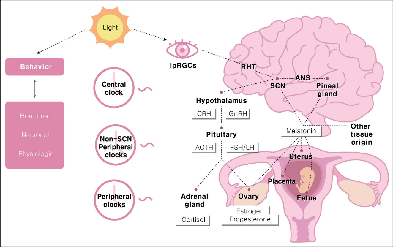
As the earth rotates, mammals encounter a 24-hour light/dark cycle. Circadian rhythm is a fundamental feature of mammalian physiology that has developed over thousands of years under continuous evolutionary pressure to survive [1,2]. Most organisms on this planet have a clock that serves to anticipate and adapt to environmental demands [3]. The most accepted concept for the mammalian circadian system is a hierarchical multi-oscillator system [4]. There is a central clock within the suprachiasmatic nucleus (SCN) of the hypothalamus, and peripheral clocks have been identified in numerous tissues, including the cerebral cortices, liver, kidney, heart, skin, and retina [5,6]. SCN neurons are circadian pacemakers with the intrinsic capacity to generate an endogenous periodicity of approximately 24 hours in isolation from other neurons [7-10].
The daily and seasonal circadian rhythms of the human body are controlled through bidirectional communication between the central and peripheral tissue clocks. The main entraining signal is light, mainly through melanopsin-containing intrinsically photosensitive retinal ganglion cells that communicate light directly to the SCN [11]. The SCN sends humoral and neuronal signals to the peripheral circadian tissue [12] and synchronizes the clocks of peripheral organs, including the female reproductive system (Figure 1).
The molecular basis of these circadian oscillations is an intracellular clock network composed of an autoregulatory transcription/translation feedback loop of molecular clock transcription regulators. In mammals, the core loop includes the transcriptional activator, brain and muscle arnt-like 1 (BMAL1) and its binding partner circadian locomotor output cycles kaput (CLOCK), including the repressors period (PER1/2/3) and cryptochrome (CRY1/2) [13,14]. PER and CRY act as potent repressors of BMAL1: CLOCK-dependent transcription in a negative feedback loop [14]. Almost all reproductive tissues have consistently been described to possess a molecular clock [15].
Successful reproduction, which requires precise timing, is crucial to the survival of a species. In the case of parturition, humans and monkeys (diurnal animals) show peaks in the middle of the night and early in the morning. In contrast, nocturnal animals such as rats and mice give birth in the afternoon [16,17]. To understand these epidemiological results, it is necessary to understand the female reproductive system in terms of the circadian rhythm. This review summarizes the circadian regulation that contributes to determining the timing of female reproduction, emphasizing the role of light and endocrine signals within the female reproductive axis.

MULTI-OSCILLATORY SYSTEM IN THE REPRODUCTIVE SYSTEM

Both the central SCN and peripheral circadian clocks play essential roles in the female reproductive system. The hypothalamic-pituitary-gonadal axis is a functional multi-oscillatory axis. The gonadal function is controlled by a collection of neurons in the hypothalamus that produces gonadotropin-releasing hormone (GnRH). GnRH is synthesized in neurons scattered throughout the preoptic area and the vascular organ of the lamina terminalis. These neurons project to the median eminence, where they release GnRH into the portal circulation in a pulsatile manner to induce proper gonadotropin secretion [18,19].
Current evidence supports the idea that circadian rhythms in kisspeptin neurons might be involved in the control of hypothalamic hormone release in rats. It is also known that other neurotransmitters and hormones, including gamma-aminobutyric acid, glutamate, neuropeptide Y, vasoactive intestinal peptide (VIP), and nitric oxide, play a role. Kisspeptin neurons are located within the hypothalamus in the anteroventral periventricular nucleus (AVPV) and arcuate nucleus. In mammals, kisspeptin neurons are found in the discrete hypothalamic nuclei. The AVPV kisspeptin neurons in particular mediate positive feedback promoting the luteinizing hormone (LH) surge. This contrasts with kisspeptin from the arcuate nucleus, which mediates negative sex steroid feedback [20-23]. GnRH neurons express a kisspeptin receptor (Kiss1R). In females, AVPV kisspeptin neurons are key to the preovulatory surges in GnRH and LH [24-27]. AVPV neurons show circadian oscillations in the expression of the clock genes PER1 and BMAL1, which lie at the core of the mammalian circadian clock [28].
Kisspeptin neurons are activated by a daily signal provided via SCN input. In rodents, environmental light cues synchronize the daily rhythm through the two main neurotransmitters released by SCN, vasopressin, and VIP [29,30]. In the late afternoon, when there are high estradiol (E2) levels exerting positive feedback on kisspeptin neurons, vasopressin activates those neurons, which release neuropeptides to the GnRH neurons, triggering the LH surge in female rodents. Furthermore, Arg-Phe amide-related peptide-3 (RFRP-3) neurons located in the dorsomedial hypothalamus, project to GnRH neurons and inhibit them. Interestingly, although both RFRP neurons and AVPV kisspeptin neurons have daily rhythms of neuronal activity, RFRP neuronal activity is observed not only in the proestrus but also the diestrus stage in rodents [31]. This indicates that RFRP neuronal activity does not depend on circulating E2 levels.

THE OVARIAN CYCLE AND THE TIMING OF OVULATION

Although temporal cues controlled by the central pacemaker (SCN) are essential for driving rhythmicity, secondary clocks located in other central structures and organs play an intrinsic role in reproduction timing. Non-SCN central clocks, such as melatonin, glucocorticoids, autonomic inputs, body temperature, and peripheral clocks, act as circadian oscillators [32].
During the human female follicular phase (metestrus-diestrus in rodents), gonadotrophs produce more follicle-stimulating hormone (FSH) than LH. This relative preponderance of FHS leads to the recruitment and development of ovarian follicles. FSH promotes follicular growth, leading to a progressive increase in E2 secretion and higher LH receptor expression in granulosa cells [33,34]. During this early phase, LH pulses occur with a high frequency (period of 1–2 hours in women). The uniform amplitude and low level of circulating E2 induce negative feedback. However, a marked surge in GnRH and LH secretion occurs, according to the positive feedback of high levels of circulating E2 during the luteal phase in women (proestrus-estrus in rodents). In this phase, the LH pulse frequency decreases to an interval of 2–6 hours with a variable amplitude [35]. A preovulatory LH surge occurs approximately every 28 days in women (4–5 days in rodents). The ovarian follicle is subdivided both anatomically and functionally into mesenchyme-derived theca cells, cuboidal androgen- and estrogen-producing cells that line the outside of the follicle, and epithelial granulosa cells that line the inside of the follicle, surround the oocyte, and primarily synthesize estrogen. Luteinization causes changes in the pattern of steroidogenic enzyme expression in both theca and granulosa cells [36]. GnRH and LH surges trigger ovulation of mature follicles within 24–48 hours in women. The LH surge also recommences oocyte meiosis while arresting granulosa cell proliferation and luteal induction. The timing of ovulation, limited to a temporal window in the afternoon of proestrus, depends on the timing of the LH surge [37,38]. Both hormonal and circadian controls of LH surge timing are necessary for ovulation. The primary role of kisspeptin neurons in reproduction is to stimulate GnRH neurons to release GnRH peptides into the hypophyseal-portal system, promoting pituitary gonadotrophs to release FSH and LH into the bloodstream [39-41].
The LH surge begins in the early morning in women [42] and diurnal (predominantly active during daylight) rodents [43]. However, in nocturnal rodents, the LH surge begins in the early afternoon [44]. Specifically, GnRH and LH surges occur at the end of the night in women [42,45] and diurnal rodents [46], whereas it occurs in the late afternoon in nocturnal rodents [47,48]. As discussed in the previous section, according to rodent studies, the SCN induces a coordinated increase in stimulatory kisspeptin via vasopressin and a decrease in the inhibitory RFRP-3 via VIP to allow a timed LH surge at the end of the resting period. For this precise time control, the core clock genes (BMAL1, CLOCK, PER1/2) and their transcripts are rhythmically expressed in the mature granulosa and luteal cells [49-51].

TIMING OF MATING AND SEASONAL REPRODUCTION

The goal of fertilization is the union of a single selected sperm nucleus with the female pronucleus within the activated oocyte. This requires appropriate mating behavior. In humans, copulation behaviors occur more frequently in the late-night, with a minor peak in the early morning [52,53]. In addition, BMAL1 contributes to maintaining neural circuits that drive pheromone-mediated mating behaviors [54]. The timing of sexual intercourse in relation to ovulation strongly influences the chances of conception. In women, the fertile window lasts approximately 6 days, ending on the day of ovulation [55].
In animals, reproductive activity is restricted to a particular time of the year to address survival environmental challenges. These seasonal reproductive cycles depend on the photoperiod, which indicates the length of the daily light phase. The cyclic melatonin production is the primary cue controlling the onset of breeding. In sheep, which are short-day breeders, changing photoperiod from longer day light to shorter day light with more periods of darkness initiates the reproductive activity. Mating occurs during increasing day lengths (spring), and the breeding season occurs during the short days of autumn and winter. The breeding seasons of hamsters are spring/summer. When female hamsters were exposed to short light period conditions, they became acyclic and anovulatory. They showed an afternoon LH surge and a small FSH increase, suggesting that they were in prolonged proestrus. Therefore, hamsters are also categorized as long day breeders [56]. The duration of elevated circulating melatonin at night depends on night length, with longer melatonin production in short-day conditions than in long-day conditions. Interestingly, when female hamsters were maintained in a long light period and injected with melatonin for several days, their estrus cycle became acyclic, and the pattern of LH and FSH secretion was similar to that found during a long dark period. Taken altogether, it is clear that melatonin plays an essential role in synchronizing seasonal reproductive activity [57].
In adult humans, the synthesis and secretion of melatonin increase shortly after the onset of darkness, with maximal levels usually observed during the middle of the night. Although melatonin is primarily secreted in the pineal gland, enzymes that convert serotonin to melatonin are also expressed in other tissues, including the reproductive tract [58]. Light-induced melatonin suppression is wavelength-dependent, with the highest response induced by short wavelengths such as blue light [59]. The response begins at the retina and is mediated by melanopsin-containing intrinsically photosensitive retinal ganglion cells [60]. Melatonin, whose receptors are present in the SCN, has important effects on neuronal firing and clock gene expression in the central pacemaker [2,61-63]. Melatonin acts at the pars tuberalis of the adenohypophysis to regulate the synthesis of thyroid-stimulating hormone (TSH). During long days, increased TSH secretion leads to higher concentrations of the thyroid hormone T3 that can activate arcuate kisspeptin. This results in GnRH-driven gonadotropin secretion and reproductive activity [34,64].

CIRCADIAN RHYTHMS IN PREGNANCY

Pregnancy, which lasts for approximately 38 weeks after conception, is a complex, multistage process that supports fetal development and optimal delivery [65,66]. For these processes, successful maternal adaptations are required, from marked central modifications in brain function to fundamental changes in reproductive, respiratory, cardiovascular, and metabolic functions [67]. Body temperature, leukocyte count, blood pressure, weight gain, rhythms of uterine contraction, blood flow, and intra-amniotic fluid pressure all follow circadian rhythms during normal pregnancy [68]. According to a recent study in mice [69], the daily activity starting time shifted earlier at the beginning of pregnancy and then back to the pre-pregnancy state approximately one week before delivery. Similarly, by using actigraphy to measure activity level, the time of sleep onset shifted earlier during the first and second trimesters and then returned to the pre-pregnancy state during the third trimester (week 28 until delivery). These results indicate that pregnancy induces changes in daily rhythms. Another more recent study [70] made similar observations also in mice that activity levels decreased during pregnancy, and activity onset was delayed. These behaviors are regulated by the circadian rhythm system.
Endocrine metabolic adaptations are required for maternal circadian rhythms during pregnancy. First, glucocorticoid hormones (cortisol in humans and corticosterone in rats and mice) are powerful mammalian hormonal axes that promote the release of energy stores to meet high fetal demands [71]. Glucocorticoid release is controlled by the hypothalamic-pituitary-adrenal axis. There is a multisynaptic neuronal connection between the SCN and autonomic preganglionic neurons that innervate the adrenal cortex, and the adrenal molecular clock regulates adrenocorticotropic hormone (ACTH) steroidogenesis in rhythmic variations [72,73]. The SCN directly and indirectly innervates corticotropinreleasing factor (CRF) neurons, which stimulates the presumed circadian rhythm of CRF secretion and downstream secretion of ACTH and cortisol [74]. ACTH and cortisol concentrations peak in the early morning, coupled with the habitual waking time according to natural light exposure. Levels decline during the daytime in pregnant humans [75-77].
Melatonin is crucial for circadian adaptation during pregnancy. As mentioned above, melatonin affects the timing of hypothalamic-pituitary-adrenal axis activation and mating in animals. Melatonin also acts as a homeostatic hormone during pregnancy, regulating several aspects of fetal physiology. During normal human pregnancy, nighttime maternal serum melatonin levels increase after 24 weeks of gestation, with significantly higher levels from 32 weeks until term [78]. Placenta-derived melatonin directly scavenges free radicals. This process reduces oxidative damage to placental tissues [79,80] and regulates the expression of antioxidant enzymes such as catalase and manganese superoxide dismutase [81]. Melatonin is thought to protect mononuclear villous cytotrophoblasts from apoptosis; therefore, they are able to continuously regenerate to fuse with and maintain a healthy syncytiotrophoblast layer [82,83]. Furthermore, maternal pineal melatonin can even pass through the placenta; therefore, it can be an important factor for entraining fetal circadian rhythms [84,85].

CIRCADIAN TIMING OF LABOR ONSET AND PARTURITION

Parturition is defined by the increasingly frequent uterine contractions accompanying cervical effacement during pregnancy that ultimately lead to the delivery of offspring [86]. Labor begins with the transition of the myometrium from a quiescent to a contractile state. This transition is accompanied by a shift in signaling from anti-inflammatory to pro-inflammatory pathways, involving chemokines (interleukin-8), cytokines (interleukin-1 and -6), and contraction-associated proteins (oxytocin receptor, connexin 43, and prostaglandin receptors) [87]. Since the parturition process requires a secure time and place, most mammals have likely adapted to selective pressures throughout evolution [88].
Parturition occurs at night or during the day depending on the temporal niche of the species. In most animals studied, it occurs immediately before or during the sleep/resting phase. For example, rats deliver predominantly during the subjective day [89-92]. Golden hamsters also deliver in the daytime [93]. Human parturition occurs preferentially at night. According to one retrospective study in the UK [94], labor that starts at night (22:00–06:00) appears to be more efficient than labor that begins during the day (10:00–18:00). Night-onset labor is more likely to result in a “normal” delivery than day-onset labor, undergoing significantly less augmentation with synthetic oxytocin and artificial rupture of membranes. In addition, more normal deliveries and fewer cesarean sections and epidurals are performed during night-onset labor. An Italian retrospective study observed a diurnal rhythm for the onset of labor, with the maximal frequency at night [95]. Similarly, the initiation of labor peaks between 24:00 and 05:00, and births follow after approximately 4 hours [96]. In the case of preterm labor, labor most commonly begins between 24:00 and 06:00 [97]. Even in twin pregnancies, a significant rhythm in the timing of contractions was noted, with 45% of deliveries occurring after labor that commenced between 24:00 and 08:00 [98]. Altogether, labor occurs preferentially during the night in diurnal species and during the day in nocturnal species.
The propensity for nighttime parturition may be related to the synergism between nocturnal increases in melatonin and oxytocin [84,86]. Oxytocin is released in a pulsatile fashion, with maximal levels in maternal plasma reached during the second and third stages of labor; paracrine interactions involving the oxytocin/oxytocin receptor system located in maternal and fetal tissues are important in the initiation of human parturition [99]. However, maternal circulating levels of oxytocin do not significantly increase during labor onset in humans. Instead, they increase more during the final expulsive stage of delivery [88]. Oxytocin receptor expression in the myometrium increases throughout pregnancy, reaching peak levels at the onset of labor. Thus, the uterus becomes gradually more sensitive to oxytocin and is most responsive for the parturition process [100,101].
Pinealectomized female rats, whose endogenous melatonin was eliminated, had normal estrous cyclicity and intact ability to conceive. However, they gave birth randomly throughout the day. Evening administration of melatonin was effective in restoring normal daytime parturition [17]. In an in-vitro study of human myometrial cells, melatonin synergistically enhanced oxytocininduced contractility via the melatonin receptor MT2R [102]. Similarly, melatonin concentration differs significantly in elective cesarean section and emergency cesarean section after induced labor. This suggests that neuroendocrine synergy between melatonin and oxytocin is possible, and that melatonin plays a key role in the onset of uterine contraction [103]. Although MTNR1A and MTNR1B melatonin receptors are expressed in the myometrium of both non-pregnant and pregnant women, MTNR1B (MT2) expression was higher in the myometrium of pregnant women in labor at term [102]. MTNR1B activates the nuclear exclusion of the androgen receptor via activation of protein kinase C [104]; this is similar to oxytocin’s mechanism of action. Therefore, it has been suggested that serum melatonin acts synergistically with oxytocin via the melatonin receptor to membrane-bound phospholipase C and protein kinase C pathways. These pathways promote the expression of the gap junction protein connexin 43 and increase uterine sensitivity to oxytocin, thereby increasing uterine contractility [105]. These findings suggest the reason for the high level of nocturnal uterine contractions that occur during lateterm human pregnancy and result in nocturnal labor.

CONCLUSION

The female reproductive system requires precise timing of processes. Rhythmicity has been demonstrated in ovulation, parturition during the resting phase, and even seasonal breeding. Animals would have needed optimal adaptation for survival and reproduction on Earth. However, recently, the environment has been significantly changing due to artificial light. It has been studied about shift work and night light exposure. It suggested that the female reproductive system could be affected by circadian disruptions. Therefore, understanding the circadian timing of the female reproductive system will be the basis for examining changes in reproductive function due to artificial light exposure.

NOTES

Funding Statement

None

Conflicts of Interest

The author has no potential conflicts of interest to disclose.

Availability of Data and Material

Data sharing not applicable to this article as no datasets were generated or analyzed during the study.

Figure 1.
Schematic model of circadian timing system in female reproduction. Light is main entraining signal in circadian timing system. Light from ipRGCs reaches the SCN through RHT. Central SCN clocks transmit the timing information to peripheral clocks along hormonal, neuronal and other physiologic pathways. Non-SCN central clocks, such as melatonin, autonomic inputs acts as circadian oscillator. HPG and HPA axis work as a functional multi-oscillatory system in female reproduction. ipRGCs, intrinsically photosensitive retinal ganglion cells; SCN, suprachiasmatic nucleus; RHT, retinohypothalamic tract; HPG, hypothalamic-pituitary-gonadal; HPA, hypothalamopituitary-adrenal; ANS, autonomic nervous system; CRH, corticotropin-releasing hormone; GnRH, gonadotropin-releasing hormone; ACTH, adrenocorticotropic hormone; FSH, follicle-stimulating hormone; LH, luteinizing hormone.
cim-2022-0017f1.jpg

REFERENCES

1. Paranjpe DA, Sharma VK. Evolution of temporal order in living organisms. J Circadian Rhythms 2005;3:7.
crossref pmid pmc
2. Gerhart-Hines Z, Lazar MA. Circadian metabolism in the light of evolution. Endocr Rev 2015;36:289–304.
crossref pmid pmc
3. Colwell CS. Linking neural activity and molecular oscillations in the SCN. Nat Rev Neurosci 2011;12:553–569.
crossref pmid pmc pdf
4. Honma S. The mammalian circadian system: a hierarchical multi-oscillator structure for generating circadian rhythm. J Physiol Sci 2018;68:207–219.
crossref pmid pmc pdf
5. Reinberg A, Bicakova-Rocher A, Nouguier J, Gorceix A, Mechkouri M, Touitou Y, et al. Circadian rhythm period in reaction time to light signals: difference between right- and left-hand side. Brain Res Cogn Brain Res 1997;6:135–140.
crossref pmid
6. Brown SA, Azzi A. Peripheral circadian oscillators in mammals. Handb Exp Pharmacol 2013;217:45–66.
crossref
7. Green DJ, Gillette R. Circadian rhythm of firing rate recorded from single cells in the rat suprachiasmatic brain slice. Brain Res 1982;245:198–200.
crossref pmid
8. Groos G, Hendriks J. Circadian rhythms in electrical discharge of rat suprachiasmatic neurones recorded in vitro. Neurosci Lett 1982;34:283–288.
crossref pmid
9. Welsh DK, Logothetis DE, Meister M, Reppert SM. Individual neurons dissociated from rat suprachiasmatic nucleus express independently phased circadian firing rhythms. Neuron 1995;14:697–706.
crossref pmid
10. Honma S, Shirakawa T, Katsuno Y, Namihira M, Honma K. Circadian periods of single suprachiasmatic neurons in rats. Neurosci Lett 1998;250:157–160.
crossref pmid
11. Foster RG, Hughes S, Peirson SN. Circadian photoentrainment in mice and humans. Biology (Basel) 2020;9:180.
crossref pmid pmc
12. Golombek DA, Rosenstein RE. Physiology of circadian entrainment. Physiol Rev 2010;90:1063–1102.
crossref pmid
13. Hastings M, O’Neill JS, Maywood ES. Circadian clocks: regulators of endocrine and metabolic rhythms. J Endocrinol 2007;195:187–198.
crossref pmid
14. Lowrey PL, Takahashi JS. Genetics of circadian rhythms in Mammalian model organisms. Adv Genet 2011;74:175–230.
crossref pmid pmc
15. Sen A, Hoffmann HM. Role of core circadian clock genes in hormone release and target tissue sensitivity in the reproductive axis. Mol Cell Endocrinol 2020;501:110655.
crossref pmid pmc
16. Ratajczak CK, Asada M, Allen GC, McMahon DG, Muglia LM, Smith D, et al. Generation of myometrium-specific Bmal1 knockout mice for parturition analysis. Reprod Fertil Dev 2012;24:759–767.
crossref pmid
17. Takayama H, Nakamura Y, Tamura H, Yamagata Y, Harada A, Nakata M, et al. Pineal gland (melatonin) affects the parturition time, but not luteal function and fetal growth, in pregnant rats. Endocr J 2003;50:37–43.
crossref pmid
18. Chryssikopoulos A, Gregoriou O, Papadias C, Loghis C. Gonadotropin ovulation induction and pregnancies in women with Kallmann’s syndrome. Gynecol Endocrinol 1998;12:103–108.
crossref pmid
19. Knobil E, Plant TM, Wildt L, Belchetz PE, Marshall G. Control of the rhesus monkey menstrual cycle: permissive role of hypothalamic gonadotropin-releasing hormone. Science 1980;207:1371–1373.
crossref pmid
20. Gu GB, Simerly RB. Projections of the sexually dimorphic anteroventral periventricular nucleus in the female rat. J Comp Neurol 1997;384:142–164.
crossref pmid
21. Petersen SL, Ottem EN, Carpenter CD. Direct and indirect regulation of gonadotropin-releasing hormone neurons by estradiol. Biol Reprod 2003;69:1771–1778.
pmid
22. Wintermantel TM, Campbell RE, Porteous R, Bock D, Gröne HJ, Todman MG, et al. Definition of estrogen receptor pathway critical for estrogen positive feedback to gonadotropin-releasing hormone neurons and fertility. Neuron 2006;52:271–280.
crossref pmid pmc
23. Glanowska KM, Venton BJ, Moenter SM. Fast scan cyclic voltammetry as a novel method for detection of real-time gonadotropin-releasing hormone release in mouse brain slices. J Neurosci 2012;32:14664–14669.
crossref pmid pmc
24. Caraty A, Franceschini I, Hoffman GE. Kisspeptin and the preovulatory gonadotrophin-releasing hormone/luteinising hormone surge in the ewe: basic aspects and potential applications in the control of ovulation. J Neuroendocrinol 2010;22:710–715.
crossref pmid
25. Kinoshita T, Caño-Delgado A, Seto H, Hiranuma S, Fujioka S, Yoshida S, et al. Binding of brassinosteroids to the extracellular domain of plant receptor kinase BRI1. Nature 2005;433:167–171.
crossref pmid
26. Smith JT, Popa SM, Clifton DK, Hoffman GE, Steiner RA. Kiss1 neurons in the forebrain as central processors for generating the preovulatory luteinizing hormone surge. J Neurosci 2006;26:6687–6694.
crossref pmid pmc
27. Watanabe Y, Uenoyama Y, Suzuki J, Takase K, Suetomi Y, Ohkura S, et al. Oestrogen-induced activation of preoptic kisspeptin neurones may be involved in the luteinising hormone surge in male and female Japanese monkeys. J Neuroendocrinol 2014;26:909–917.
crossref pmid pdf
28. Smarr BL, Gile JJ, de la Iglesia HO. Oestrogen-independent circadian clock gene expression in the anteroventral periventricular nucleus in female rats: possible role as an integrator for circadian and ovarian signals timing the luteinising hormone surge. J Neuroendocrinol 2013;25:1273–1279.
crossref pmid pmc pdf
29. Francl JM, Kaur G, Glass JD. Regulation of vasoactive intestinal polypeptide release in the suprachiasmatic nucleus circadian clock. Neuroreport 2010;21:1055–1059.
crossref pmid pmc
30. Kalsbeek A, Buijs RM, Engelmann M, Wotjak CT, Landgraf R. In vivo measurement of a diurnal variation in vasopressin release in the rat suprachiasmatic nucleus. Brain Res 1995;682:75–82.
crossref pmid
31. Henningsen JB, Ancel C, Mikkelsen JD, Gauer F, Simonneaux V. Roles of RFRP-3 in the daily and seasonal regulation of reproductive activity in female Syrian hamsters. Endocrinology 2017;158:652–663.
crossref pmid
32. de Assis LVM, Oster H. The circadian clock and metabolic homeostasis: entangled networks. Cell Mol Life Sci 2021;78:4563–4587.
crossref pmid pmc pdf
33. Hillier SG. Current concepts of the roles of follicle stimulating hormone and luteinizing hormone in folliculogenesis. Hum Reprod 1994;9:188–191.
crossref pmid
34. Simonneaux V. A kiss to drive rhythms in reproduction. Eur J Neurosci 2020;51:509–530.
crossref pmid pdf
35. Kaiser UB. Gonadotrophin hormones. In: Melmed S. editors. The pituitory (3rd ed). Cambridge, MA: Elsevier, Academic Press, 2011, p. 205–260.

36. Stouffer RL, Hennebold JD. Structure, function, and regulation of the corpus luteum. In: Neill JD. editors. Knobil and Neill’s physiology of reproduction (4th ed). Cambridge, MA: Elsevier, Academic Press, 2015, p. 1023–1076.

37. Kriegsfeld LJ, Silver R. The regulation of neuroendocrine function: timing is everything. Horm Behav 2006;49:557–574.
crossref pmid pmc
38. Moenter SM, DeFazio AR, Pitts GR, Nunemaker CS. Mechanisms underlying episodic gonadotropin-releasing hormone secretion. Front Neuroendocrinol 2003;24:79–93.
crossref pmid
39. Irwig MS, Fraley GS, Smith JT, Acohido BV, Popa SM, Cunningham MJ, et al. Kisspeptin activation of gonadotropin releasing hormone neurons and regulation of KiSS-1 mRNA in the male rat. Neuroendocrinology 2004;80:264–272.
crossref pmid pdf
40. Han SK, Gottsch ML, Lee KJ, Popa SM, Smith JT, Jakawich SK, et al. Activation of gonadotropin-releasing hormone neurons by kisspeptin as a neuroendocrine switch for the onset of puberty. J Neurosci 2005;25:11349–11356.
crossref pmid pmc
41. Messager S, Chatzidaki EE, Ma D, Hendrick AG, Zahn D, Dixon J, et al. Kisspeptin directly stimulates gonadotropin-releasing hormone release via G protein-coupled receptor 54. Proc Natl Acad Sci U S A 2005;102:1761–1766.
crossref pmid pmc
42. Kerdelhué B, Brown S, Lenoir V, Queenan JT Jr, Jones GS, Scholler R, et al. Timing of initiation of the preovulatory luteinizing hormone surge and its relationship with the circadian cortisol rhythm in the human. Neuroendocrinology 2002;75:158–163.
crossref pmid pdf
43. Mahoney MM, Sisk C, Ross HE, Smale L. Circadian regulation of gonadotropin-releasing hormone neurons and the preovulatory surge in luteinizing hormone in the diurnal rodent, Arvicanthis niloticus, and in a nocturnal rodent, Rattus norvegicus. Biol Reprod 2004;70:1049–1054.
pmid
44. Bronson FH, Vom Saal FS. Control of the preovulatory release of luteinizing hormone by steroids in the mouse. Endocrinology 1979;104:1247–1255.
crossref pmid
45. Cahill DJ, Wardle PG, Harlow CR, Hull MG. Onset of the preovulatory luteinizing hormone surge: diurnal timing and critical follicular prerequisites. Fertil Steril 1998;70:56–59.
crossref pmid
46. McElhinny TL, Sisk CL, Holekamp KE, Smale L. A morning surge in plasma luteinizing hormone coincides with elevated Fos expression in gonadotropin-releasing hormone-immunoreactive neurons in the diurnal rodent, Arvicanthis niloticus. Biol Reprod 1999;61:1115–1122.
pmid
47. Legan SJ, Karsch FJ. A daily signal for the LH surge in the rat. Endocrinology 1975;96:57–62.
crossref pmid
48. Moline ML, Albers HE, Todd RB, Moore-Ede MC. Light-dark entrainment of proestrous LH surges and circadian locomotor activity in female hamsters. Horm Behav 1981;15:451–458.
crossref pmid
49. Chu G, Yoshida K, Narahara S, Uchikawa M, Kawamura M, Yamauchi N, et al. Alterations of circadian clockworks during differentiation and apoptosis of rat ovarian cells. Chronobiol Int 2011;28:477–487.
crossref pmid
50. Chu G, Misawa I, Chen H, Yamauchi N, Shigeyoshi Y, Hashimoto S, et al. Contribution of FSH and triiodothyronine to the development of circadian clocks during granulosa cell maturation. Am J Physiol Endocrinol Metab 2012;302:E645–E653.
crossref pmid
51. He PJ, Hirata M, Yamauchi N, Hashimoto S, Hattori MA. Gonadotropic regulation of circadian clockwork in rat granulosa cells. Mol Cell Biochem 2007;302:111–118.
crossref pmid pdf
52. Palmer JD, Udry JR, Morris NM. Diurnal and weekly, but no lunar rhythms in humans copulation. Hum Biol 1982;54:111–121.
pmid
53. Refinetti R. Time for sex: nycthemeral distribution of human sexual behavior. J Circadian Rhythms 2005;3:4.
crossref pmid pmc
54. Schoeller EL, Clark DD, Dey S, Cao NV, Semaan SJ, Chao LW, et al. Bmal1 is required for normal reproductive behaviors in male mice. Endocrinology 2016;157:4914–4929.
crossref pmid pmc
55. Wilcox AJ, Weinberg CR, Baird DD. Timing of sexual intercourse in relation to ovulation. Effects on the probability of conception, survival of the pregnancy, and sex of the baby. N Engl J Med 1995;333:1517–1521.
crossref pmid
56. Reiter RJ. Pineal control of a seasonal reproductive rhythm in male golden hamsters exposed to natural daylight and temperature. Endocrinology 1973;92:423–430.
crossref pmid
57. Bartness TJ, Powers JB, Hastings MH, Bittman EL, Goldman BD. The timed infusion paradigm for melatonin delivery: what has it taught us about the melatonin signal, its reception, and the photoperiodic control of seasonal responses? J Pineal Res 1993;15:161–190.
crossref pmid
58. Acuña-Castroviejo D, Escames G, Venegas C, Díaz-Casado ME, Lima-Cabello E, López LC, et al. Extrapineal melatonin: sources, regulation, and potential functions. Cell Mol Life Sci 2014;71:2997–3025.
crossref pmid pdf
59. Rüger M, St Hilaire MA, Brainard GC, Khalsa SB, Kronauer RE, Czeisler CA, et al. Human phase response curve to a single 6.5 h pulse of shortwavelength light. J Physiol 2013;591:353–363.
crossref pmid pmc
60. Hattar S, Liao HW, Takao M, Berson DM, Yau KW. Melanopsin-containing retinal ganglion cells: architecture, projections, and intrinsic photosensitivity. Science 2002;295:1065–1070.
crossref pmid pmc
61. Agez L, Laurent V, Pévet P, Masson-Pévet M, Gauer F. Melatonin affects nuclear orphan receptors mRNA in the rat suprachiasmatic nuclei. Neuroscience 2007;144:522–530.
crossref pmid
62. Gillette MU, McArthur AJ. Circadian actions of melatonin at the suprachiasmatic nucleus. Behav Brain Res 1995;73:135–139.
crossref
63. McArthur AJ, Gillette MU, Prosser RA. Melatonin directly resets the rat suprachiasmatic circadian clock in vitro. Brain Res 1991;565:158–161.
crossref pmid
64. Dardente H. Melatonin-dependent timing of seasonal reproduction by the pars tuberalis: pivotal roles for long daylengths and thyroid hormones. J Neuroendocrinol 2012;24:249–266.
crossref pmid
65. Smith R. Parturition. N Engl J Med 2007;356:271–283.
crossref pmid
66. Cha J, Sun X, Dey SK. Mechanisms of implantation: strategies for successful pregnancy. Nat Med 2012;18:1754–1767.
crossref pmid pmc pdf
67. Mark PJ, Crew RC, Wharfe MD, Waddell BJ. Rhythmic three-part harmony: the complex interaction of maternal, placental and fetal circadian systems. J Biol Rhythms 2017;32:534–549.
crossref pmid pdf
68. Valenzuela FJ, Vera J, Venegas C, Pino F, Lagunas C. Circadian system and melatonin hormone: risk factors for complications during pregnancy. Obstet Gynecol Int 2015;2015:825802.
crossref pmid pmc pdf
69. Martin-Fairey CA, Zhao P, Wan L, Roenneberg T, Fay J, Ma X, et al. Pregnancy induces an earlier chronotype in both mice and women. J Biol Rhythms 2019;34:323–331.
crossref pmid pmc pdf
70. Yaw AM, Duong TV, Nguyen D, Hoffmann HM. Circadian rhythms in the mouse reproductive axis during the estrous cycle and pregnancy. J Neurosci Res 2021;99:294–308.
crossref pmid pmc pdf
71. Atkinson HC, Waddell BJ. The hypothalamic-pituitary-adrenal axis in rat pregnancy and lactation: circadian variation and interrelationship of plasma adrenocorticotropin and corticosterone. Endocrinology 1995;136:512–520.
crossref pmid
72. Spencer RL, Chun LE, Hartsock MJ, Woodruff ER. Glucocorticoid hormones are both a major circadian signal and major stress signal: how this shared signal contributes to a dynamic relationship between the circadian and stress systems. Front Neuroendocrinol 2018;49:52–71.
crossref pmid
73. Oster H, Damerow S, Kiessling S, Jakubcakova V, Abraham D, Tian J, et al. The circadian rhythm of glucocorticoids is regulated by a gating mechanism residing in the adrenal cortical clock. Cell Metab 2006;4:163–173.
crossref pmid
74. Kalsbeek A, van der Spek R, Lei J, Endert E, Buijs RM, Fliers E. Circadian rhythms in the hypothalamo-pituitary-adrenal (HPA) axis. Mol Cell Endocrinol 2012;349:20–29.
crossref pmid
75. Patrick J, Challis J, Campbell K, Carmichael L, Natale R, Richardson B. Circadian rhythms in maternal plasma cortisol and estriol concentrations at 30 to 31, 34 to 35, and 38 to 39 weeks’ gestational age. Am J Obstet Gynecol 1980;136:325–334.
crossref pmid
76. Cousins L, Rigg L, Hollingsworth D, Meis P, Halberg F, Brink G, et al. Qualitative and quantitative assessment of the circadian rhythm of cortisol in pregnancy. Am J Obstet Gynecol 1983;145:411–416.
crossref pmid
77. Mastorakos G, Ilias I. Maternal and fetal hypothalamic-pituitary-adrenal axes during pregnancy and postpartum. Ann N Y Acad Sci 2003;997:136–149.
crossref pmid
78. Nakamura Y, Tamura H, Kashida S, Takayama H, Yamagata Y, Karube A, et al. Changes of serum melatonin level and its relationship to feto-placental unit during pregnancy. J Pineal Res 2001;30:29–33.
crossref pmid pdf
79. Soliman A, Lacasse AA, Lanoix D, Sagrillo-Fagundes L, Boulard V, Vaillancourt C. Placental melatonin system is present throughout pregnancy and regulates villous trophoblast differentiation. J Pineal Res 2015;59:38–46.
crossref pmid pdf
80. Lanoix D, Beghdadi H, Lafond J, Vaillancourt C. Human placental trophoblasts synthesize melatonin and express its receptors. J Pineal Res 2008;45:50–60.
crossref pmid
81. Richter HG, Hansell JA, Raut S, Giussani DA. Melatonin improves placental efficiency and birth weight and increases the placental expression of antioxidant enzymes in undernourished pregnancy. J Pineal Res 2009;46:357–364.
crossref pmid
82. Vaillancourt C, Lanoix D, Le Bellego F, Daoud G, Lafond J. Involvement of MAPK signalling in human villous trophoblast differentiation. Mini Rev Med Chem 2009;9:962–973.
crossref pmid
83. Lanoix D, Guérin P, Vaillancourt C. Placental melatonin production and melatonin receptor expression are altered in preeclampsia: new insights into the role of this hormone in pregnancy. J Pineal Res 2012;53:417–425.
crossref pmid
84. Reiter RJ, Tan DX, Korkmaz A, Rosales-Corral SA. Melatonin and stable circadian rhythms optimize maternal, placental and fetal physiology. Hum Reprod Update 2014;20:293–307.
crossref pmid
85. Okatani Y, Okamoto K, Hayashi K, Wakatsuki A, Tamura S, Sagara Y. Maternal-fetal transfer of melatonin in pregnant women near term. J Pineal Res 1998;25:129–134.
crossref pmid
86. Olcese J, Lozier S, Paradise C. Melatonin and the circadian timing of human parturition. Reprod Sci 2013;20:168–174.
crossref pmid pdf
87. Romero R, Dey SK, Fisher SJ. Preterm labor: one syndrome, many causes. Science 2014;345:760–765.
crossref pmid pmc
88. Olcese J. Circadian aspects of mammalian parturition: a review. Mol Cell Endocrinol 2012;349:62–67.
crossref pmid
89. Plaut SM, Grota LJ, Ader R, Graham CW 3rd. Effects of handling and the light-dark cycle on time of parturition in the rat. Lab Anim Care 1970;20:447–453.
pmid
90. Boer K, Lincoln DW, Swaab DF. Effects of electrical stimulation of the neurohypophysis on labour in the rat. J Endocrinol 1975;65:163–176.
crossref pmid
91. Lincoln DW, Porter DG. Timing of the photoperiod and the hour of birth in rats. Nature 1976;260:780–781.
crossref pmid pdf
92. Rowland DL, Wagonblast AL, Dykstra TA. Timing of parturition in the rat: an analysis of successive births. Chronobiologia 1991;18:31–38.
pmid
93. Siegel HI, Greenwald GS. Prepartum onset of maternal behavior in hamsters and the effects of estrogen and progesterone. Horm Behav 1975;6:237–245.
crossref pmid
94. Kanwar S, Rabindran R, Lindow SW. Delivery outcomes after day and night onset of labour. J Perinat Med 2015;43:729–733.
crossref pmid
95. Cagnacci A, Soldani R, Melis GB, Volpe A. Diurnal rhythms of labor and delivery in women: modulation by parity and seasons. Am J Obstet Gynecol 1998;178(1 Pt 1): 140–145.
crossref pmid
96. Serón-Ferré M, Ducsay CA, Valenzuela GJ. Circadian rhythms during pregnancy. Endocr Rev 1993;14:594–609.
crossref pmid
97. Lindow SW, Jha RR, Thompson JW. 24 hour rhythm to the onset of preterm labour. BJOG 2000;107:1145–1148.
crossref pmid
98. Rabindran R, Kanwar S, Lindow SW. 24-hour rhythm to the onset of term and preterm labour in twin pregnancies. BJOG 2010;117:1656–1657.
crossref pmid
99. Smith JG, Merrill DC. Oxytocin for induction of labor. Clin Obstet Gynecol 2006;49:594–608.
crossref pmid
100. Kimura T, Takemura M, Nomura S, Nobunaga T, Kubota Y, Inoue T, et al. Expression of oxytocin receptor in human pregnant myometrium. Endocrinology 1996;137:780–785.
crossref pmid
101. Fuchs AR, Fuchs F, Husslein P, Soloff MS. Oxytocin receptors in the human uterus during pregnancy and parturition. Am J Obstet Gynecol 1984;150:734–741.
crossref pmid
102. Sharkey JT, Puttaramu R, Word RA, Olcese J. Melatonin synergizes with oxytocin to enhance contractility of human myometrial smooth muscle cells. J Clin Endocrinol Metab 2009;94:421–427.
crossref pmid pmc
103. Bagci S, Berner AL, Reinsberg J, Gast AS, Zur B, Welzing L, et al. Melatonin concentration in umbilical cord blood depends on mode of delivery. Early Hum Dev 2012;88:369–373.
crossref pmid
104. Sampson SR, Lupowitz Z, Braiman L, Zisapel N. Role of protein kinase Calpha in melatonin signal transduction. Mol Cell Endocrinol 2006;252:82–87.
pmid
105. McCarthy R, Jungheim ES, Fay JC, Bates K, Herzog ED, England SK. Riding the rhythm of melatonin through pregnancy to deliver on time. Front Endocrinol (Lausanne) 2019;10:616.
crossref pmid pmc
TOOLS
Share :
Facebook Twitter Linked In Google+ Line it
METRICS Graph View
  • 0 Crossref
  •   Scopus 
  • 2,201 View
  • 51 Download
Related articles in Chronobiol Med


ABOUT
ARTICLE CATEGORY

Browse all articles >

BROWSE ARTICLES
EDITORIAL POLICIES
FOR CONTRIBUTORS
Editorial Office
RN1611, 725, Suseo-Dong, Gangnam-Gu, Seoul 06367, Republic of Korea
Tel: +82-2-445-1611    Fax: +82-2-445-1633    E-mail: editor@chronobiologyinmedicine.org                

Copyright © 2024 by Korean Academy of Sleep Medicine. All rights reserved.

Developed in M2PI

Close layer
prev next